Fig. 1.
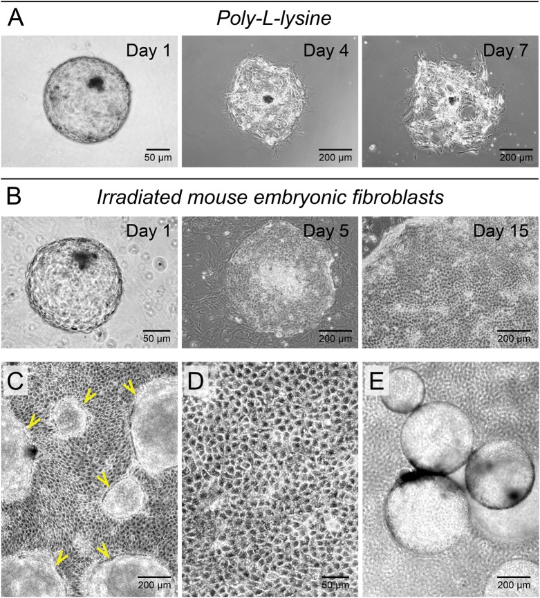
Mouse embryonic fibroblasts support attachment and growth of bovine blastocyst-derived trophoblasts. (A) Poly-L-lysine coated surfaces did not support bovine blastocyst attachment and trophoblast outgrowths. Of the blastocysts that attached, cells failed to expand and rapidly disintegrated. (B) Irradiated mouse embryonic fibroblast feeders (MEFs) allowed for blastocyst attachment and proliferation of the trophectoderm leading to colony formation. (C) Trophoblast colonies grew with limited basal attachments as sheets and formed numerous surface outpocketings (arrowheads) over time. (D) Proliferating trophoblast cells formed a characteristic polygonal cell sheet with prominent cell adhesions and resolvable cytoplasmic elements within. (E) As a result of pinch-offs from surface outpocketings, fluid-filled hollow trophoblast spheres analogous to the blastocyst-trophectoderm organization were frequently released from trophoblast colonies in culture.

