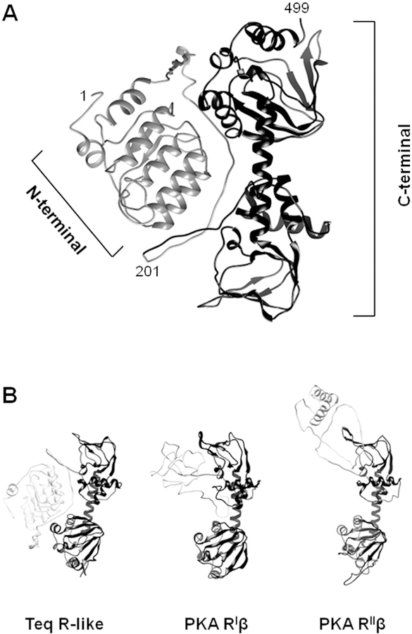
Fig. 7. Structural model of the T. equiperdum R-like protein.
A, The Phyre2 bioinformatics tool [38] was employed to predict the structure of the T. equiperdum R-like protein. Ribbon diagram structures of the modeled N-terminal (residues 1–201) and C-terminal (residues 202e499) regions are shown in light grey and dark grey, respectively. B, The model retrieved for the trypanosome protein is compared to those retrieved for PKA human RIβ (PDB ID: 4DIN [22]) and rat RIIβ (PDB ID: 3TNQ [61]). N-terminal and C-terminal regions are shown in light grey and dark grey, respectively.

