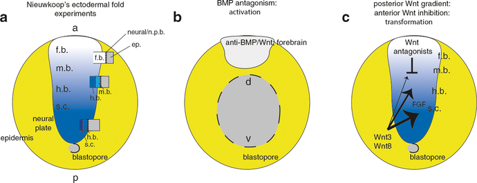
Fig. 12.
Models for anteroposterior axis patterning in vertebrates. (a) Depiction of Nieuwkoop’s ectodermal fold implantation experiments (Nieuwkoop 1952; Nieuwkoop and Nigtevecht 1954). Dorsal posterior view of a neurula stage amphibian embryo; neural fate is represented as a gradient from light-to-dark with darker color indicating more posterior fates; the epidermis is yellow. The implanted folds are shown as boxes, divided to show the approximately position of induced neural fates. Each fold is characterized by a distal epidermal portion (ep.) bounded by general neural/neural plate border (activated tissue); this is followed proximally by graded neural fates, reflecting the hypothesized influence of a transforming gradient (as opposed to a distinct inducer at each AP level). (b) Molecular interpretation of Nieuwkoop’s model. In the gastrula, neural induction is accomplished by BMP antagonism, which induces neural tissue with forebrain character. (c) During later gastrulation, the expression of Wnts directly induces posterior fates in anterior neural-fated tissue in a dose-dependent fashion. FGF signaling is required in a permissive role. Wnt antagonists expressed in the anterior mesendoderm limit the extent of Wnt signaling and the anterior remains forebrain

