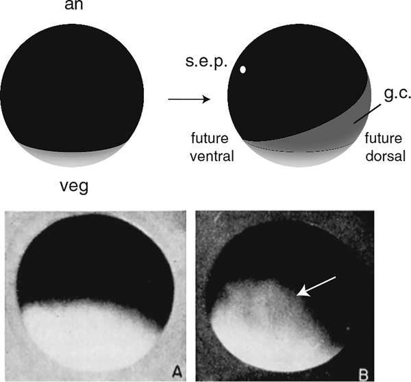
Fig. 2.
Gray crescent formation in amphibians. Top panel, diagram of an amphibian egg (e.g., Rana) before (left) and after fertilization (right). The heavily pigmented animal pole (an) and the paler vegetal pole (veg) are indicated. After fertilization, corticocytoplasmic movements opposite to the sperm entry point (s.e.p.) result in the appearance of the gray crescent (g.c.) on the prospective dorsal side. Bottom panel, images of a Rana egg at fertilization (a), and at 20 min post-fertilization, showing the gray crescent (b; dorsal view, arrow). Bottom panel reproduced from Rugh (1951)

