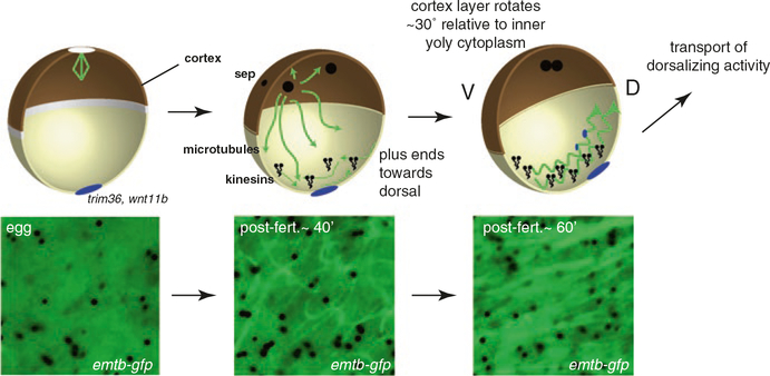
Fig. 3.
Events of cortical rotation in Xenopus. Microtubules are disassembled during oocyte maturation, and are absent from the egg cortex (left panels). Certain RNAs are localized to the vegetal cortex during oogenesis (blue) and encode proteins critical for cortical rotation and dorsalization (e.g., trim36, wnt11b). After fertilization, the incoming sperm pronucleus and associated centrosome initiate astral microtubule assembly. Cortical microtubule assembly also begins, forming a network by 40 min post-fertilization. A shear zone forms and microtubules associate with the yolky cytoplasmic core (not shown) and cortical rotation begins, under the action of kinesin-like proteins (kinesins). Relative cortical movement occurs dorsally, possibly the result of nudging by ventrally positioned astral microtubules, and rapidly orients microtubule plus ends dorsally (Olson et al. 2015) (middle panel). Microtubule assembly and organization becomes robust by 60 min post-fertilization and full cortical rotation commences, continuing until first cleavage. Rapid transport of dorsalizing activity occurs along parallel microtubule arrays using kinesin-like motors (right panel). The corresponding bottom panels show live images of microtubules labeled with Enconsin microtubule-binding domain tagged GFP (EMTB-GFP), showing progressive assembly and alignment during cortical rotation (Olson et al. 2015)

