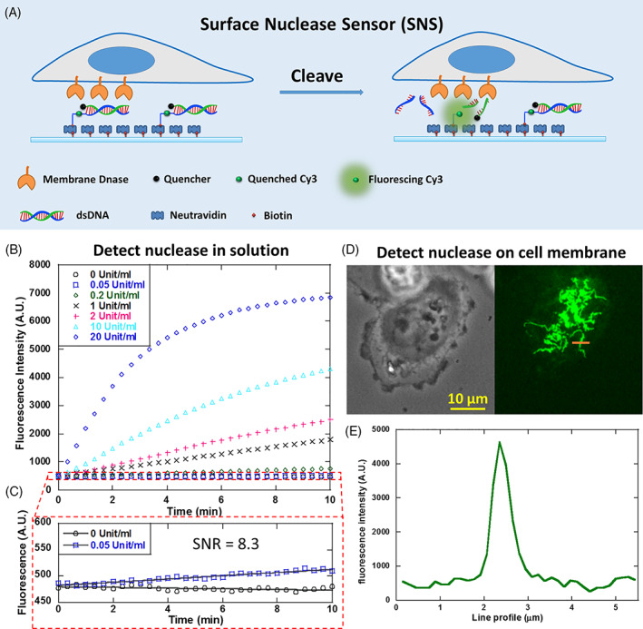
Figure 1.

Developing surface‐tethered nuclease sensor (SNS) for the in situ mapping of membrane‐bound nuclease (MN) activity on the cell membrane. (A) SNS is a fluorophore (here Cy3) conjugated with a biotin and a quencher‐labeled dsDNA. The fluorophore is freed from quenching when the dsDNA is degraded by MN, thus reporting the MN activity by fluorescence on site. (B) SNS‐coated surface reported DNase I in solution at a series of concentrations. (C) The detection limit for soluble DNase I by SNS is calibrated to be 0.01 unit/mL (rounding 0.05/8.3‐0.01). (D) An SNS‐coated surface reported highly organized MN activity on the ventral membrane of adherent MDA‐MB‐231 cells. (E) The structure feature of the SNS pattern is finer than 1 μm
