
Figure 6.
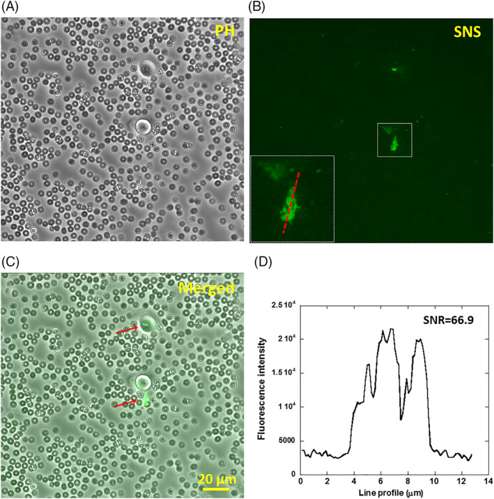
Detecting breast cancer cells in blood sample using SNS. (A‐C) Canine whole blood diluted by 10 fold and mixed with breast cancer cells (MDA‐MB‐231), mimicking circulating tumor cells in blood, was plated on a surface coated with SNS and fibronectin. SNS signal were acquired after 1 hour of cell incubation. The number ratio between MDA‐MB‐231 cells and red blood cells is 1:106. (D) The SNS signal caused by MN activity has a high contrast to the dark background with a signal‐to‐noise ratio of 66.9, making it feasible to differentiate cancer cells from a large background of blood cells