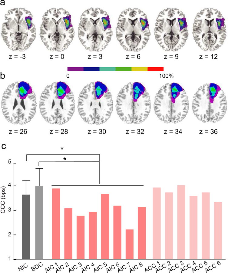
Figure 7. Impaired CCC in patients with lesions in the AIC.
Lesion reconstruction for patients with unilateral lesion in anterior insular cortex (AIC group) and (b) patients with unilateral lesion in anterior cingulate cortex (ACC group). All lesions were mapped on right hemisphere. Colors indicate the percentage of the overlap of lesions across patients. (c) Estimated CCC of participants in different groups. NIC (dark grey bar): neurological intact control. BDC (light grey bar): brain damage control, referring to patients with lesion in regions outside the cognitive control network. Error bars indicate the standard deviation. Pink bar: each patient in the AIC group (left AIC lesion: 2 and 7; right AIC lesion: AIC 1, 3, 4, 5, 6, and 8). Light pink bar: each patient in the ACC group (left ACC lesion: ACC 3, 5, and 6; right ACC lesion: ACC 1, 2, and 4). *: p < .05.

