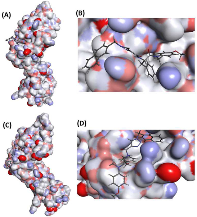
Fig. 4.
Representative structures of the first (A) and second (C) conformational clusters and their corresponding expanded views of 13m binding to the first (B) and second clusters (D). Protein part of the representative structures was illustrated as surface to show the binding site and conformations.

