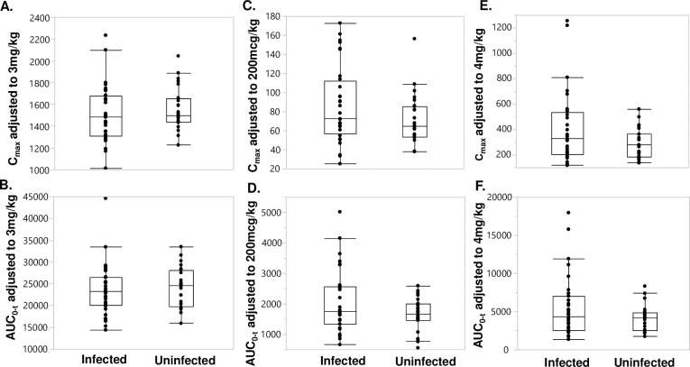
Fig 3.
Distribution of dose adjusted Cmax and AUC0-t of DEC (A and B), IVM (C and D), and ALB-OX (E and F) stratified by LF infection status. The median, 25th, and 75th quartiles and 95% CI are shown. Significance was assessed with the Kruskal-Wallis test and all P values were >0.05. There were 32 LF-infected and 24 uninfected participants.

