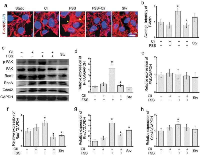
Figure 4.

Inhibition of integrin attenuated FSS-induced cytoskeleton rearrangement. (a) HepG2 cells were loaded with FSS at 1 dyn/cm2 for 0.5 h, with or without treatment of 0.5 µM Cliengitide (Cli) for 6 h prior to FSS application. Immunostaining of F-actin (Red), nuclei (Blue) were performed. The yellow arrow indicated the stress fibers. (b) The average intensity of F-actin was analyzed from at least 10 random field (4*104 μm2) (n = 3). (c) HepG2 cells were loaded with FSS at 1 dyn/cm2 for 0.5 h, with or without treatment of 0.5 µM Cli for 6 h prior to FSS application. Lysates were probed with antibodies as indicated. (d-g) Quantification of protein expressions in (c).
*p < 0.05 vs. Static; #p < 0.05 vs. FSS.
