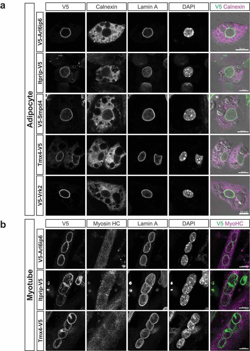
Figure 6.

Immunofluorescence analysis of the localization of target proteins in adipocytes and myocytes. (a) C3H cells stably transduced with the indicated constructs were differentiated into adipocytes and co-labeled with antibodies to the V5 epitope tag, calnexin and lamins A. DNA staining (DAPI) and a merge of V5 and calnexin labeling is shown in the right panels. (b) C2C12 myoblasts that were stably transduced with the indicated constructs were differentiated into myotubes and labeled as in (a). Scale bars, 10μm.
