. 2019 May 3;8:e44760. doi: 10.7554/eLife.44760

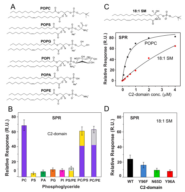
Figure 4. cPLA2α C2-domain binding affinity for phosphoglyceride and sphingomyelin (SM) vesicles.

(A) Phosphoglyceride structural formulas. (B) Relative affinities of the C2-domain (1 μM) for different phosphoglycerides obtained by SPR. Molar ratios for PS/PE, PC/PS and PC/PE mixed composition vesicles are 7:3. (see Figure 4—figure supplement 1). (C) SPR binding isotherms showing C2-domain equilibrium adsorption to immobilized POPC or 18:1-SM vesicles as a function of protein concentration (see Figure 4—figure supplement 2 for additional information). (D) Effect of C2-domain mutations (1 μM) on binding to 18:1 SM obtained by SPR (see 'Materials and methods' for other details).

Figure 4—source data 1. cPLA2α C2-domain binding affinity for phosphoglyceride and sphingomyelin (SM) vesicles.

Figure 4.

Figure 4—figure supplement 1. Assessment of C2-domain binding to different phosphoglycerides.

Figure 4—figure supplement 1.

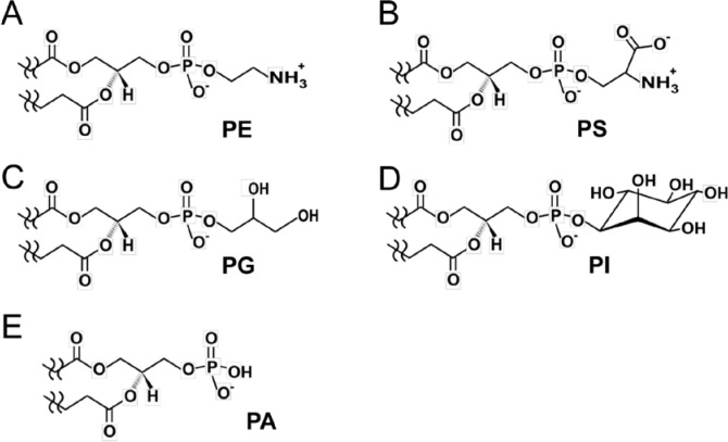
Head group chemical formulas of various non-PC membrane phosphoglycerides are (A) phosphatidylethanolamine, PE; (B) phosphatidylserine, PS; (C) phosphatidylglycerol, PG; (D) phosphatidylinositol, PI; and (E) phosphatidic acid, PA. All of the phosphoglycerol-based lipids have the same backbone topology but different head groups that define C2-domain binding specificity. Our findings reveal that the π-cation interaction is critical for binding to PC. PE (A) has a primary ammonium group replacing the –N+(CH3)3 group in PC. Yet, earlier FRET data (Nalefski et al., 1998) and our SPR data (Figure 4B) indicate relatively weak binding of cPLA2α C2-domain to PE. The electrostatic potential difference and diminished van der Waals contacts with Ala94, His62, and Asn64 could account for the binding affinity decrease for PE compared to PC. PS has a seryl group (B) replacing the –N+(CH3)3 group in PC. Although the primary ammonium group in the seryl group would seem to be a candidate for undergoing π-cation interaction with Tyr96, binding by C2-domain is weak (Figure 4B) suggesting steric clashing of the seryl carboxylate group with CBL residues. PG (C) and PI (D) are not suitable for interaction with cPLA2α C2-domain due to lack of an ammonium group and steric clashing by their bulky head groups. PA (E) has only a phosphoryl moiety as its head group, which promotes weak interaction. Our SPR binding data showing much weaker binding of these phosphoglyerides compared to PC (Figure 4B) are consistent with previous findings obtained using other techniques (Mosior et al., 1998; Nalefski et al., 1998; Six and Dennis, 2003).
Figure 4—figure supplement 2. The concentration- and time-dependence of C2-domain adsorption/desorption to/from immobilized POPC and 18:1 SM vesicles measured by SPR.
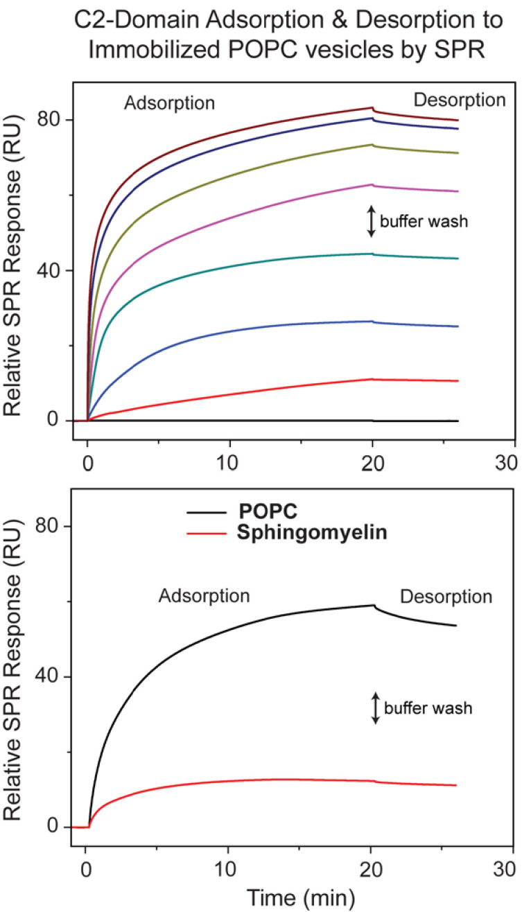
Figure 4—figure supplement 2.

(Upper panel) SPR data showing cPLA2α C2-domain adsorption/desorption to/from immobilized POPC vesicles at varying protein concentrations (0.1, 0.2, 0.4, 0.6, 1, 2, and 4 μM; bottom to top) and 5 μl/min flow rate. The two-headed arrow indicates switch to buffer containing no protein. The same approach was used to assess C2-domain adsorption to immobilized 18:1-SM vesicles shown in Figure 4C and 4D. The Kd values calculated from the binding isotherms for C2-domain and the point mutants are reported in Table 1. (Lower panel) SPR data showing the time-dependence for cPLA2α C2-domain adsorption/desorption to/from immobilized 18:1 SM vesicles at 0.6 μM protein concentration.