
Fig. 6. MSC encapsulation in vitro within PdBT cross-linked gels.
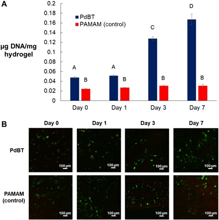
(A) Double-stranded DNA content of live cells normalized to wet hydrogel mass is shown at point of fabrication (0 days) and over the course of 7 days of in vitro culture for hydrogels cross-linked by either PdBT or an established PAMAM cross-linker. Data are reported as means ± SD for a sample size of n = 3. Different letters A to D indicate statistically significant differences between time points. (B) Representative LIVE/DEAD images are shown for cross-sectional slices of hydrogels at each time point. Green and red staining indicates live and dead cells, respectively.