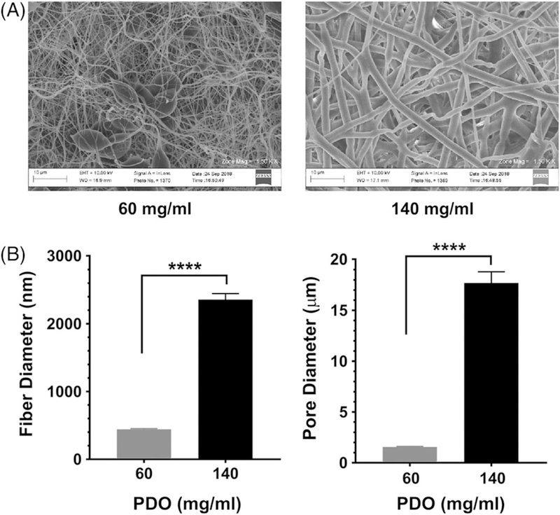
FIGURE 1.

Electrospun scaffold structure varies with PDO concentration. (A) Polydioxanone scaffolds of different polymer concentrations were electrospun, yielding scaffolds of different morphologies, as confirmed by SEM. (B) Electron micrographs were used to measure fiber and pore diameters of the scaffolds, calculated with ImageJ. Data shown are mean ± SEM from 25 to 60 measurements. ****, p < 0.0001.
