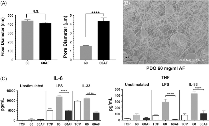
FIGURE 4.

I ncreasing pore size of 60 mg/mL electrospun scaffolds suppresses IL-6 and TNF production. (A) Fiber and pore diameters of the indicated scaffolds were calculated from SEM images shown at right. Data shown are mean ± SEM of 60 measurements. (B) Representative electromicrographs of a 60AF scaffold at two magnifications, showing regions of increased porosity. (C) BMMC were seeded on fibronectin-coated 60 and 60 AF electrospun scaffolds for 48 h and then activated with LPS (1 μg/mL) or IL-33 (100 ng/mL). Supernatants were collected 16 h later and analyzed via ELISA for IL-6 and TNF. Results are presented as mean ± SEM of nine samples from three independent experiments. ****, p < 0.0001.
