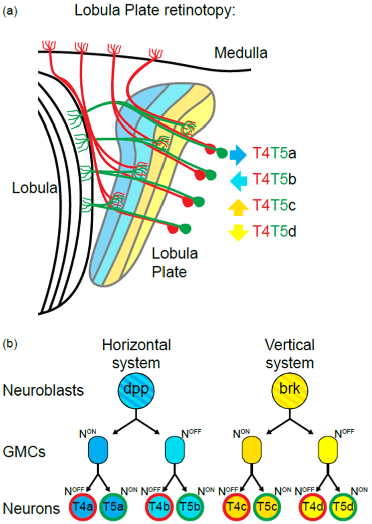
Figure 4:

Retinotopic formation in the lobula plate:
(a) Schematic of the lobula plate and the 4 subtypes of T4 (in red) and T5 neurons (in green). Each subtype targets to a different layer of the lobula plate neuropile (color coded for their response to motion direction, arrow on the side
(b) Two neuroblasts give rise to the 4 T4s and T5s of a single column. Both go through two rounds of Notch mediated asymmetric decision and give rise to the 2 T4s and T5s of either the horizontal system for the Dpp+ neuroblast or of the vertical system for the Brk+ neuroblast.
