
Figure 2 –
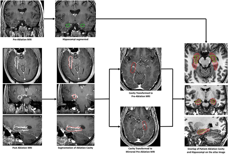
Workflow for image-processing. For each patient, the ablation cavity was manually segmented according to previously-described methods.13 Preoperative and postoperative images along with the manually-segmented ablation cavity were normalized to a common reference space.15 Once completed for the entire cohort, a critical population-based analysis of ablation volumes and locations could be performed.