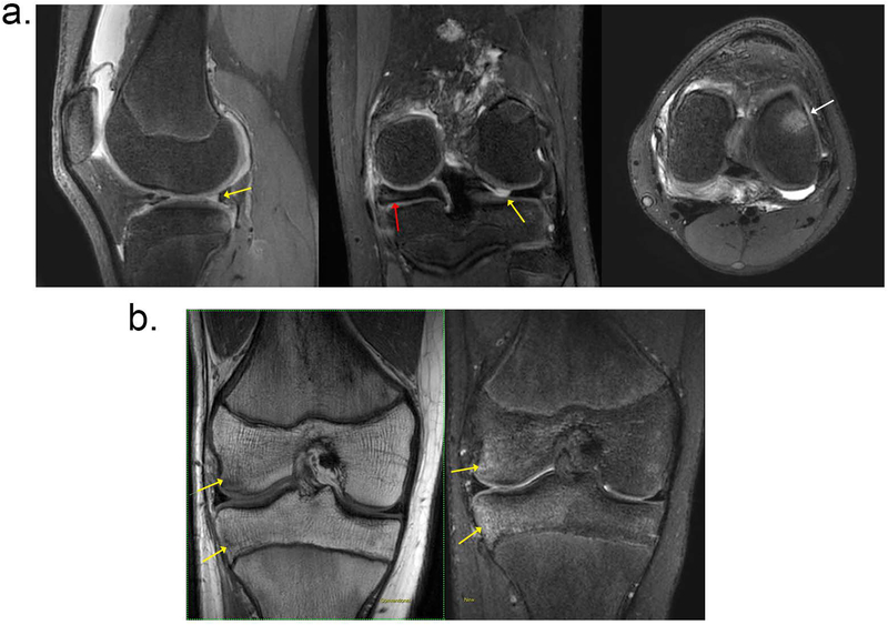
Figure 2.
Two patients with clinical indication of meniscal tear. (a) A 13-year-old female patient evaluated for internal derangement of the left knee. T2Sh images reformatted into (left) sagittal PD, (middle) coronal T2, and (right) axial intermediate weighting. Clinical suspicion of lateral meniscal tear was confirmed with MRI (yellow arrows). Additional related findings were medial discoid meniscus (red arrow) and bone marrow edema (white arrow). (b) Patient presented with knee pain and clinical suspicion of meniscal tear; (left) coronal nonfat-suppressed T1, and (right) T2Sh reformatted to coronal T2 weighted images are shown. Note the bone bruise, marked by the yellow arrows.

