Figure 7.
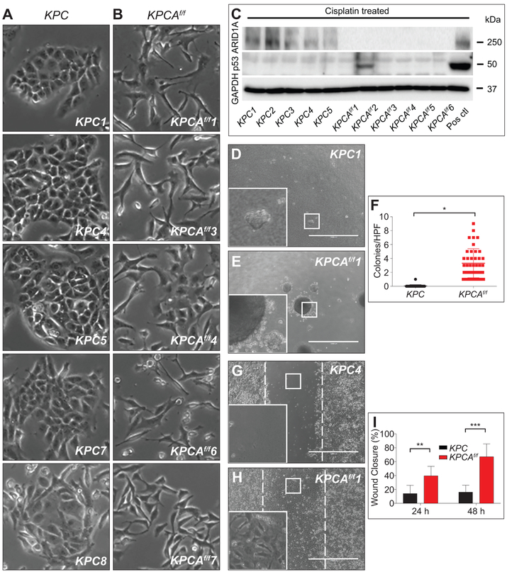
Arid1a loss increases anchorage-independent growth and migration. (A) KPC tumour cell lines (n=5) demonstrating epithelial morphology compared to (B) KPCAf/f tumour cell lines (n=5) showing mesenchymal morphology. (C) Western blot of protein lysates from KPC and KPCAf/f cell lines shows absence of ARID1A as expected and absence of P53 induction following cisplatin treatment in all but one KPCAf/f cell lines. GAPDH served as a loading control. Positive control is an Arid1a/p53 intact PDAC line (Pos ctl). (D-F) Clonogenic assay and quantification showing (E,F) KPCAf/f cell lines (n=2) form greater numbers of colonies in soft agar compared to (D,F) KPC (n=2), *p=8.5×10⁻¹³. Colonies ≥125 μm were counted. (G-I) Wound healing assay and quantification demonstrate that (H,I) KPCAf/f cell lines (n=2) have increased ability to fill the wound area compared to (G,I) KPC (n=2) at (I) 24 and 48 hours, **p=6.45×10⁻⁸, ***p=4.81×10⁻¹⁵. Analysis by TScratch. Bar=1000 μm for (D,E,G,H).

