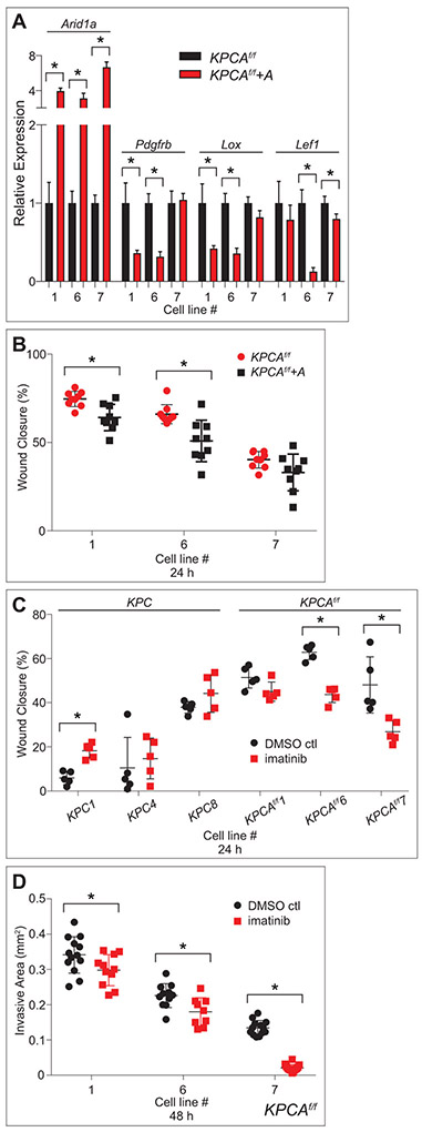
Figure 9.
Re-expression of Arid1a and inhibition of PDGFRb partially abrogate the phenotypes of KPCAf/f cell lines. (A) qPCR data showing re-expression (KPCAf/f+A) and controls (KPCAf/f) in three KPCAf/f cell lines demonstrate significantly increased expression of Arid1a mRNA in all three lines, and significantly decreased expression of Pdgfrb, Lox, and Lef1 in two of three lines each. (B) Quantification of wound healing assay in three KPCAf/f cell lines with (KPCAf/f+A) and without (KPCAf/f) Arid1a re-expression demonstrates that KPCAf/f+A have decreased ability to fill the wound area compared to controls in two of three cell lines tested. (C) Quantification of wound healing assay in three KPC and KPCAf/f cell lines treated with imatinib (1 μM) and DMSO control demonstrates that imatinib has no effect on most KPC cell lines tested while imatinib-treated KPCAf/f cell lines have decreased ability to fill the wound area compared to controls in two of three lines tested. (B,C) All measured at 24 hours, *p<0.05, analysis by TScratch. (D) Quantification of cancer cell spheroid invasion assay in three KPCAf/f cell lines treated with imatinib (1 μM) and DMSO control reveals decreased total area invaded by imatinib-treated KPCAf/f cells leaving the spheroid in all three cell lines tested. All measured at 48 hours, *p<0.05, analysis by ImageJ.

