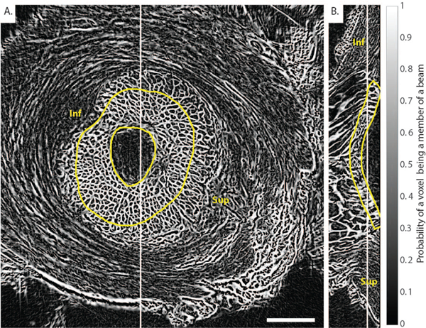
Fig. 4. Frangi’s vesselness filter for connective tissue detection applied to the PC μCT scan of Pig B at 6mmHg.

A) Transverse slice and B) sagittal slice. A modified version of Frangi’s vesselness filter was applied to enhance plate-like objects in the 3D image volume shown in Fig. 3. The image is a probability map that each voxel is a member of a beam. The method clearly identified the beam-like connective tissue constituents of the LC but did not evenly segment the non-trabecular structure of the sclera, as expected. Inf and Sup indicate inferior (Ventral) and superior (Dorsal) orientations, respectively. Scale bar = 1.25 mm.
