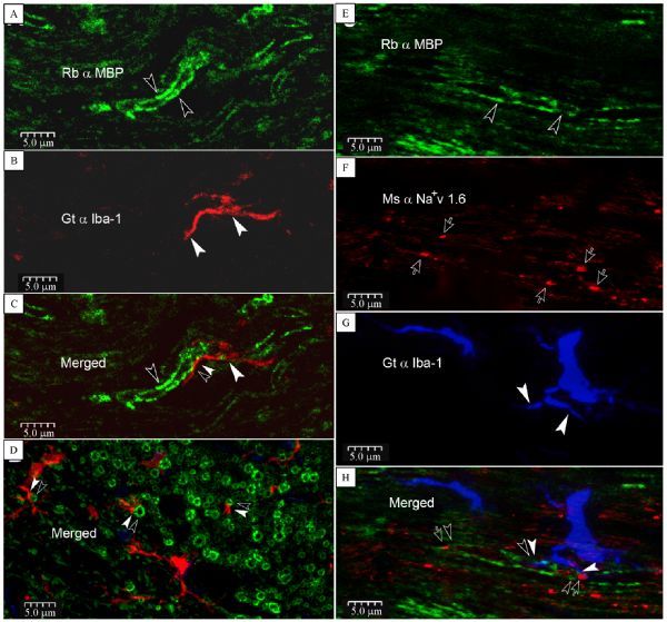
Fig.1. Direct contacts of microglia pseudopodia with inter-, para-nodal myelin sheath and probably Ranvier’s node (RN).

A¨CC: Iba-1 positive microglia pseudopodium (arrowheads) directly contacts onto myelin basic protein (MBP) labeled myelin sheath (opened arrowheads) in the corpus callosum (CC, paired arrowheads). D: contacts (paired arrowheads) between microglia pseudopodia and myelin sheathes viewed in cross section. E¨CH: a single microglia with its pseudopodia (arrowheads) in close apposition upon a RN (opened arrows) and its para- and internodal myelin sheath (opened arrowheads). Merged panel shows Iba-1 positive process contacts on a MBP labeled internodal (paired down-point arrowheads) myelin sheath. Meanwhile, the other pseudopodium seems to contact with a RN and its paranodal apparatus (upper-point opened arrow and arrowhead, plus a left-point arrowhead). This image is constructed from 3 scanned layers to show an entire single microglia with both soma and processes.
