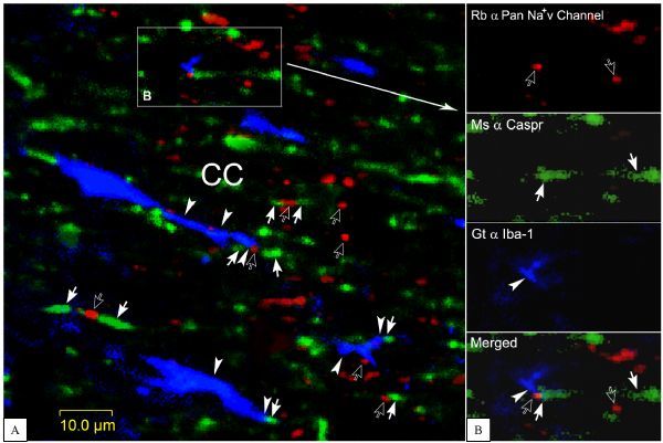
Fig.3. Verification of microglia pseudopodia contact with RNs and paranodal domains.

A: three typical structures are viewed herein. 1, Na+v channel positive RNs (opened arrows) nested in-between of Caspr 1 labeled paranodal domains (arrows and an opened arrow together); 2, microglia pseudopodia (arrowheads) contact on RNs lining with paranodal domains (arrow, arrowhead and opened arrow together); 3, microglia pseudopodia are tightly apposite upon paranodal domains (arrowhead and arrow together). B: from a framed area in A, showing a labeled microglia pseudopodium immediately contacts a RN and its paranodal domain (arrowhead, opened arrow and arrow together in merged panel). This image was constructed from 3 laser scanned layers. CC, corpus callosum.
