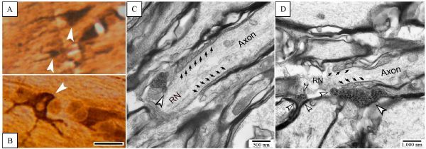
Fig.4. Ultrastructural view of microglia pseudopodia contacts on RNs.

A and B: Iba-1 immunohistochemically labeled, osmicated and plate-embedded sections observed under bright-field microscope, on which the labeled microglia and pseudopodia (arrowheads) could be seen clearly. Scale bar = 15 ¦̭. C and D: ultrathin sections cut from A or B, exhibiting ends of microglia pseudopodia (opened arrowheads) direct contact on naked axolemma of RN area. Apposition of microglia pseudopodium upon paranodal myelin sheath is also viewed (D), but there seems to be a tiny gap (about 5¨C10 nm) between them. Aligned arrows point towards spiral loops of paranodal apparatus.
