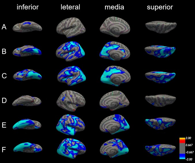
Figure 3.
Differences in cortical thickness among patients with Alzheimer’s disease (AD), patients with amnestic mild cognitive impairment (aMCI) and normal controls (NC). The comparison is made from four directions (inferior, lateral, medial and superior) in the left hemisphere (A, B and C) and right hemisphere (D, E and F), respectively. (A) and (D) Cortical thickness did not significantly differ between patients with aMCI and NC in both left and right hemisphere. (B) and (E) The blue areas showed significantly reduced cortical thickness in patients with aMCI compared with NC. (C) and (F) The blue areas showed significantly reduced cortical thickness in patients with AD compared with aMCI.

