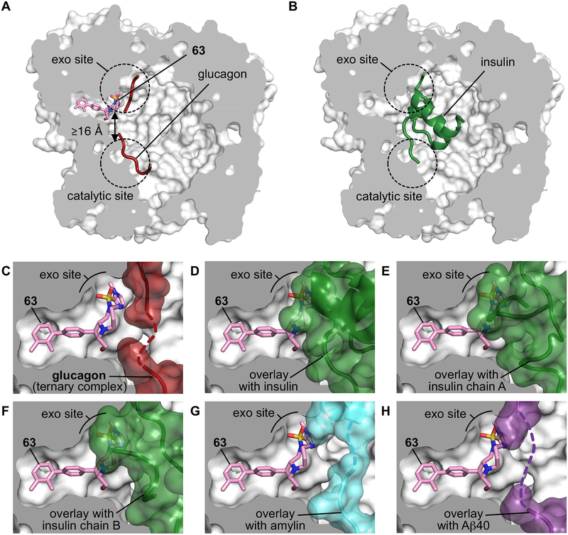
Fig. 4 |. Structural basis for substrate-selective small-molecule inhibition of IDE.
(a–b) X-ray co-crystal structures of 63 and glucagon bound to IDE as a ternary complex IDE•63•glucagon (a, PDB ID 6EDS, 3.18 Å resolution) compared to the previously reported structure of insulin-bound IDE (PDB ID 2WBY, b)40. (c) View of the exo site in the IDE•63•glucagon co-crystal structure showing the space-filling model of glucagon (red). The dashed red line represents disordered residues in the central section of glucagon (see Supplementary Figures 3–4). (d–h) Matching views of the exo-site of IDE bound by 63 in which glucagon has been cloaked, and shown instead with superimposed substrates from published IDE-substrate co-crystal structures40,42. (d) Shows the superimposition with partially folded insulin bound to IDE (green, PDB ID 2WBY), (e) with unfolded insulin α-chain from cryo-EM insulin•IDE structure (green; PDB ID 6BFC), (f) with unfolded insulin β-chain from cryo-EM insulin•IDE structure (green; PDB ID 6B3Q), (g) with amylin bound to IDE (PDB ID 2G48; cyan surface), and (h) with Aβ(1–40) bound to IDE (PDB ID 2G47; purple surface)40,42. Supplementary Figure 3 includes complementary analyses for the X-ray co-crystal structures of IDE•37 and IDE•63 co-crystalized without glucagon. See the Supplementary Video associated with this figure.

