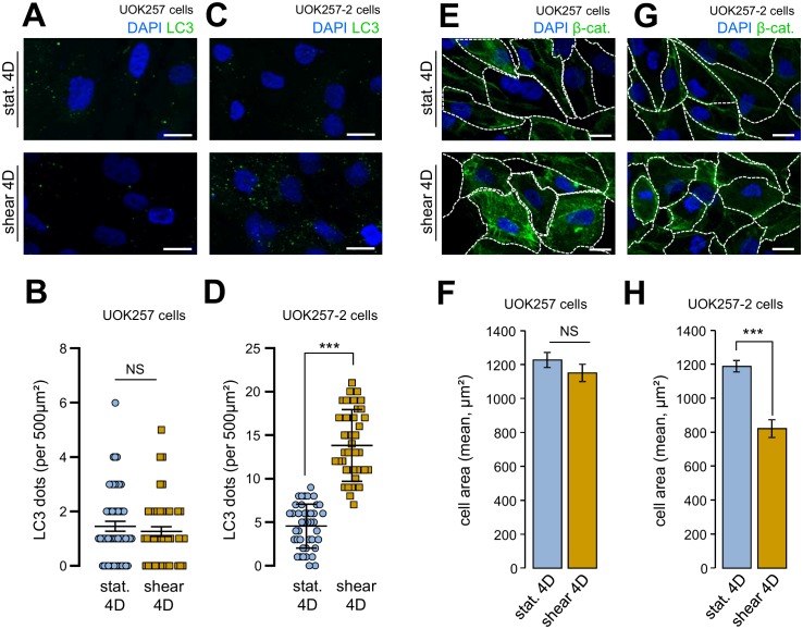
Figure 5. FIGURE 5: Shear-stress-induced autophagy and cell size regulation are abolished in FLCN-null cells.
UOK 257 FLCN-null cells and UOK 257-2 FLCN restored cells were cultured on microslides and then subjected to fluid flow for 4 days (shear) or not (static 4D). Cells were fixed, labeled with DAPI, immunostained for LC3 (A, C) or labeled with DAPI and immunostained for β-catenin to reveal cell boarder (E, G) and then analyzed by fluorescence microscopy. (B, D) LC3 dots were quantified from experiments shown in (A) (UOK 257cells) and (C) (UOK 257-2 cells). (F, H) cells areas were quantified from experiments shown in (E) (UOK 257cells) and (G) (UOK 257-2 cells). Scale bars in (A, C, E and F) = 10μm.

