Figure 3.
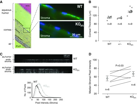
Nbce1b/c-null (KOb/c) mice exhibit corneal swelling and increased corneal opacity. (A) Light micrograph of a hematoxylin and eosin stained wild-type (WT) mouse eye section showing the approximate locations highlighted in the accompanying fluorescence microscopy images of the corneas from WT and KOb/c mice. Note the absence of Nbce1 immunoreactivity (using the anti-Slc4a4 antibody) from the corneal endothelia of KOb/c mice (representative of images gathered from eye sections from three pairs of mice). (B) The in vivo (left eye) corneal thickness of WT mice (two males, six females), together with data gathered from their heterozygous (+/−; four males, two females) and KOb/c (four males, five females) littermates. *P<0.05: significantly different from WT thickness according to ANOVA with Tukey post-hoc analysis. (C) Representative z-axis projections of confocal reflection micrographs of the corneas of freshly enucleated WT and KOb/c mouse eyes (the endothelium is visible as a bright band, the epithelium could not be imaged because of its proximity to the highly reflective coverslip) together with a distribution plot of the pixel intensities of the stroma. (D) The median stromal pixel intensities for a larger number of WT and KOb/c mice.

