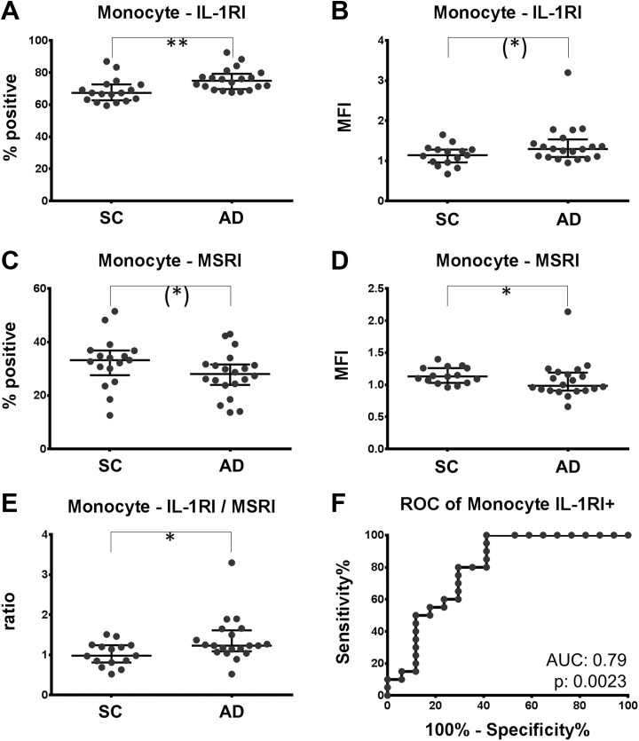
Figure 1.
Expression of IL-1RI is increased; expression of MSRI is decreased on monocytes of patients with AD. PBMC of patients with AD and SC was labeled with antibodies directed against IL-1RI and MSRI. A and C, Fraction of monocytes staining positive for IL-1RI and MSRI. B and D, The mean fluorescence intensity (MFI) of the IL-1RI and the MSRI on monocytes are depicted. The ratio of IL-1RI (MFI)/MSRI (MFI) comparing the 2 populations is plotted in (E). F, The receiver operating characteristic curve (ROC) for the discrimination of patients with and without AD by the fraction of IL-1RI-positive monocytes. Trend: *P < .10; significance: *P < .05; **P < .01. AD indicates Alzheimer disease; IL-1RI, interleukin 1 receptor subtype I; MSRI, macrophage scavenger receptor I; PBMC, peripheral blood mononuclear cell; SC, symptomatic controls.

