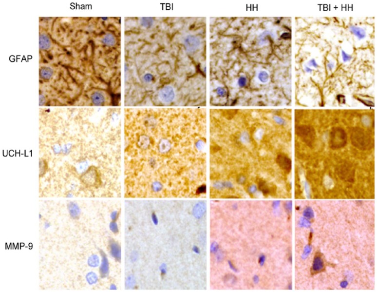
Figure 8.
Immunohistochemical staining patterns of GFAP, UCH-L1, and MMP-9 proteins in each group: Sham, TBI, HH, and TBI+HH in the neocortex.
All magnifications are at ×40. GFAP proteins form filament network in cytoskeleton and staining is less intensive in TBI, HH, and TBI+HH groups compared with Sham group. UCH-L1 proteins have diffuse and granulated staining with more intensive staining in the TBI, HH, and TBI+HH groups compared with sham group. For MMP-9, stainings are almost inexistant for Sham and TBI groups and are marked and diffuse for HH and TBI+HH groups. GFAP indicates glial fibrillary acidic protein; HH, hypoxia/hypotension group; MMP-9, matrix metalloproteinase 9; Sham, Sham group; TBI, traumatic brain injury group; TBI+HH, traumatic brain injury and hypoxia/hypotension group; UCH-L1, ubiquitin carboxy-terminal hydrolase-L1.

