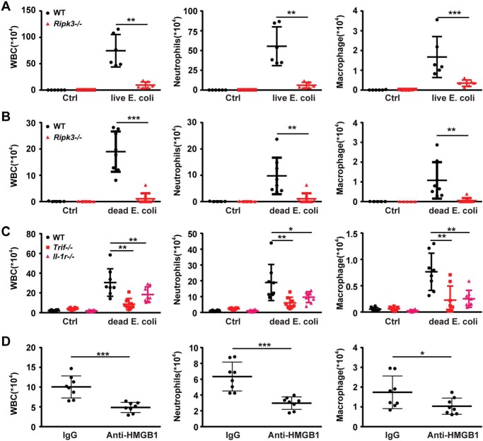
Figure 7.
Loss of RIPK3 attenuates inflammation induced by dead E. coli and HMGB1. A, air-pouch lavage fluid was collected from WT and Ripk3−/− mice following injection with live E. coli for analysis of infiltrated white blood cell (WBC) counts by microscope (left) and neutrophils (middle) and macrophages (right) by flow cytometry. B, air-pouch lavage fluid was collected from WT and Ripk3−/− mice following injection with heat-killed E. coli for analysis of infiltrated white blood cell counts by microscope (left) and neutrophils (middle) and macrophages (right) by flow cytometry. C, WT, TrifLps/Lps2 mice and Il-1R−/− mice were selected for the same experiment as B, and leukocytes (left), neutrophils (middle), and macrophages (right) numbers are shown. D, air-pouch inflammatory infiltration was induced in the absence or presence of HMGB1-neutralizing or normal control IgGs. Then air-pouch lavage fluid was collected, and leukocytes (left), neutrophils (middle), and macrophages (right) numbers are shown. Circles represent individual mice. *, p < 0.05; **, p < 0.01; ***, p < 0.001. Graphs show the mean ± S.D. from three independent experiments.

