Figure 2.
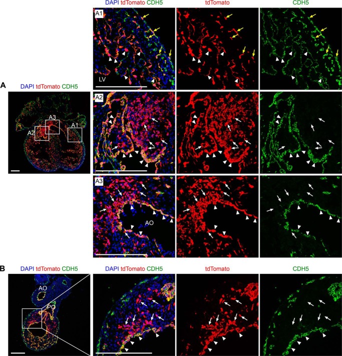
Nfatc1–Dre labels endocardium, coronary endothelial cells, and endocardial-derived cushion MCs. A and B, immunostaining for tdTomato and CDH5 on heart sections from Nfatc1–Dre;R26–rsr–tdTomato at E13.5. The boxed regions in the left panels are magnified and split channels in the right panels as indicated. The yellow arrows in A1 indicate tdTomato+CDH5+ coronary endothelial cells. The white arrows in A2 indicate tdTomato+CDH5− MCs in atrioventricular cushion. The white arrows in A3 indicate tdTomato+CDH5− MCs in aortic outflow tract (AO). The white arrows in B indicate tdTomato+CDH5− MCs in pulmonary outflow tract (PO). The arrowheads in A and B indicate tdTomato+CDH5+ endocardial cells. DAPI, 4′,6′-diamino-2-phenylindole. Scale bars, 200 μm. Each picture is representative of three individual embryonic samples.

