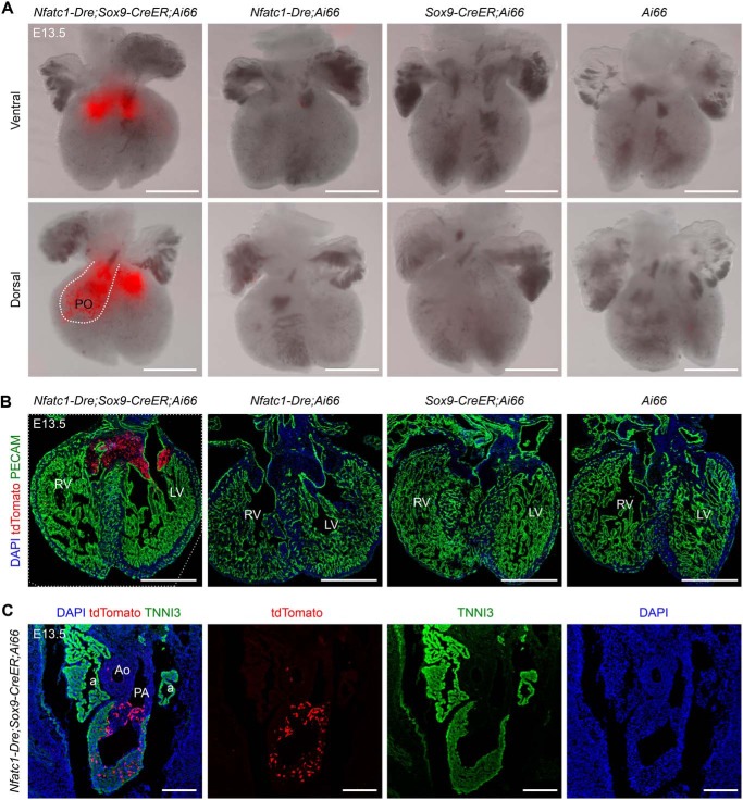
Figure 4.
Endocardial-derived MCs in the pulmonary outflow tract invade the myocardium at E13.5. A, whole-mount fluorescence view of the hearts with indicated genotypes, which were administered with tamoxifen at E9.5 and harvested at E13.5. The dotted line indicates pulmonary outflow tract (PO). B, immunostaining for tdTomato and PECAM on embryonic hearts with indicated genotypes at E13.5. C, immunostaining for tdTomato and TNNI3 on embryonic sections from Nfatc1–Dre;Sox9–CreER;Ai66 at E13.5. RV, right ventricle; LV, left ventricle; a, atrium; Ao, aorta; PA, pulmonary artery; DAPI, 4′,6′-diamino-2-phenylindole. Scale bars, 500 μm. Each picture is representative of three individual mouse samples.

