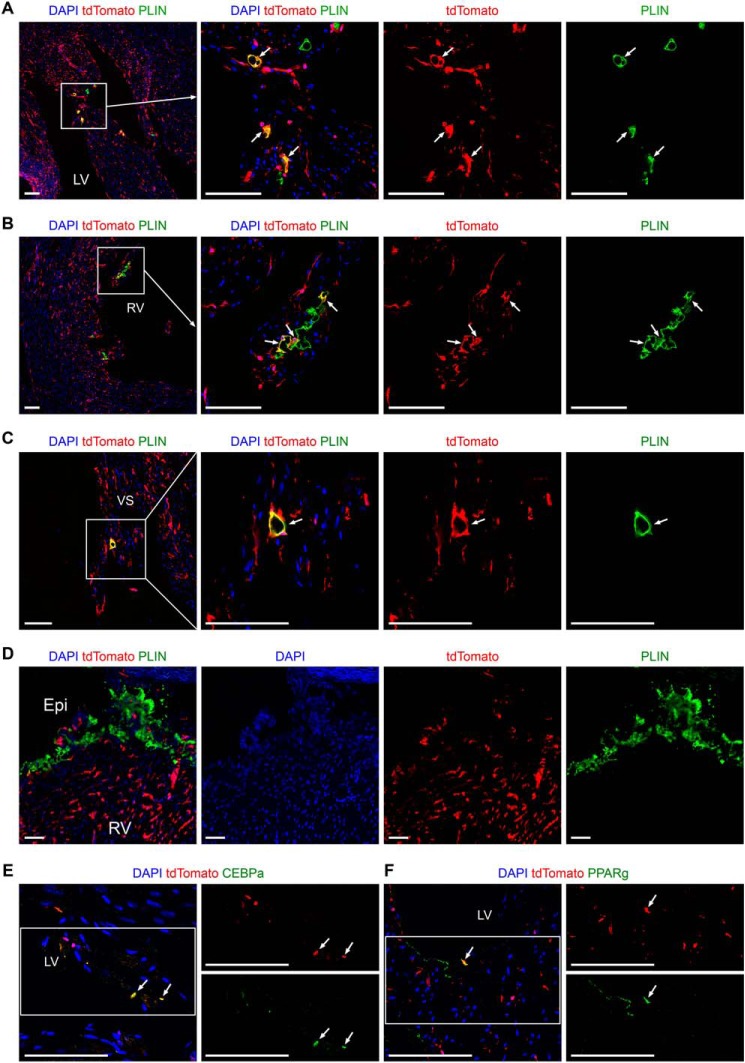
Figure 8.
Intersectional lineage tracing shows the contribution of endocardial-derived MCs to intramyocardial adipocytes in adult hearts. A–D, immunostaining for tdTomato and PLIN on heart sections from adult Nfatc1–Dre;Sox9–CreER;Ai66, which were treated with tamoxifen at E9.5. tdTomato+ adipocytes are identified in LV (A), RV (B), and VS (C), but not in subepicardium (D). The boxed regions in the left panels are magnified and split channels in the right panels. The arrows indicate tdTomato+PLIN+ intramyocardial adipocytes. E and F, immunostaining for tdTomato and CEBPα or PPARγ on heart sections from adult Nfatc1–Dre;Sox9–CreER;Ai66. The arrows indicate tdTomato+CEBPα+ or tdTomato+PPARγ+ cells. The boxed regions in the left panel are magnified and split channels in the right panels. LV, left ventricle; RV, right ventricle; VS, ventricular septum; Epi, epicardium. Scale bars, 100 μm. Each picture is representative of three individual mouse samples.

