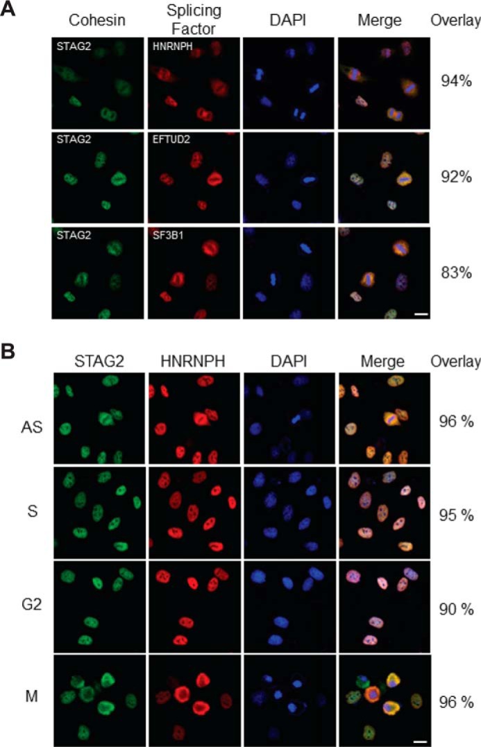
Figure 3.

Co-localization of splicing factors with cohesin. A, proliferating HeLa cells were fixed, permeabilized, and double-stained with antibodies to cohesin (STAG2) and three different interacting splicing factors (HNRNPH, EFTUD2, and SF3B1). Imaging was performed on a Leica TCS SP8 confocal laser-scanning microscope, and co-localization was quantified using ImageJ software. For technical details of immunofluorescence and co-localization analysis, see “Experimental procedures.” B, HeLa cells were treated with DMSO vehicle alone (AS), hydroxyurea (S), RO-3306 (G2), and nocodazole (M) for 24 h to arrest cells at S, G2, and M phases of the cell cycle, respectively. Cells were then fixed, permeabilized, and double-stained with antibodies to cohesin (STAG2) and a representative interacting splicing factor (HNRNPH). Microscopy and co-localization analysis were performed as in A. DAPI, 4′,6-diamidino-2-phenylindole. Bar = 10 μm.
