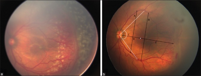
Figure 1.
(a) Technique of barrage laser. Left eye of a 34-week post menstrual age baby showing zone 2 posterior stage 3 ROP with plus disease. Posterior barrage laser spots are visible in between the tortuous vessels. (b) Image analysis: Fovea is represented by the white dot. A - Inter-arcade distance at 2 disc diameter from the temporal edge of disc. B - Inter-arcade distance at 4 disc diameter from the temporal edge of the disc. C – Angle subtended by the temporal vessels from the fovea at the center of the disc. D - Disc–fovea distance. E - Fovea–ridge distance. D + E - Disc–ridge distance

