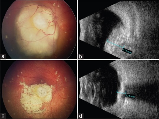
Figure 2.

(a) Fundus photograph of a 10-month-old Caucasian infant showing solitary exophytic macular retinoblastoma with focal subretinal seeds and subretinal fluid in the right eye (OD), classified as group C. (b) B-scan ultrasonography confirming a calcified intraocular measuring 7.11 mm in thickness. After receiving three sessions of IAC, (c) the tumor is completely regressed (Type 1) with resolution of subretinal fluid and (d) the tumor has reduced to 3.71 mm thickness
