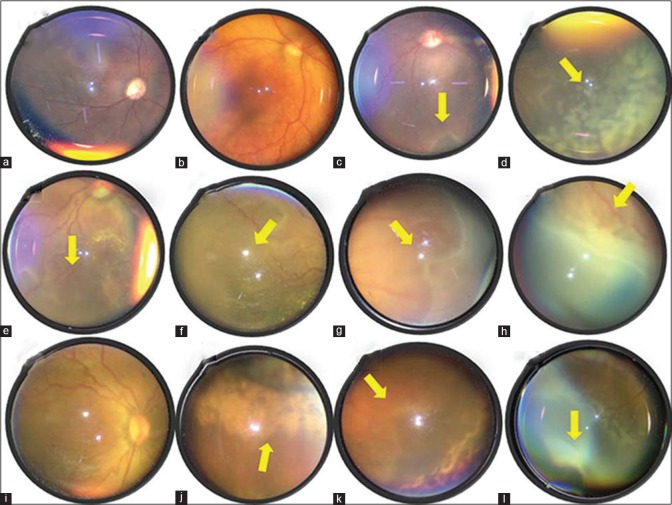
Figure 3.
Various retinopathy of prematurity (ROP) presentations – (a) glare due to mid-dilated pupil, (b) wide-field view of a normal retina, (c) plus disease with ridge (arrow) in zone 1, (d) laser marks (arrow), (e) aggressive posterior ROP in zone 1, (f) demarcation between vascular and avascular retina (arrow), (g) ridge (arrow) in zone 2, (h) stage 3 fibrovascular proliferation in zone 2 (arrow), (i) dilated tortuous vessels in all four quadrants s/o plus disease, (j) laser scar marks in periphery, (k) skip area between normal retina and laser marks (arrow), (l) ora serrata nicely seen with scleral depression

