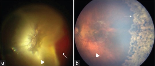
Figure 5.

(a) Right eye developed vitreous hemorrhage (arrow) and retinal detachment (arrowhead) during follow-up period. (b) Left eye shows regressed neovascularization with scatter laser marks (arrow) with attached retina (arrowhead)

(a) Right eye developed vitreous hemorrhage (arrow) and retinal detachment (arrowhead) during follow-up period. (b) Left eye shows regressed neovascularization with scatter laser marks (arrow) with attached retina (arrowhead)