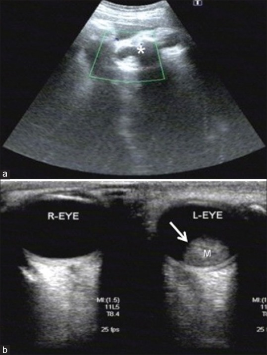
Figure 1.

(a) Obstetric ultrasound scan at 39 weeks gestation; coronal image of fetal orbit shows a hyperechoic intraocular lesion on the left side located in the retina measuring 15 × 15 × 12 mm. (white astrix) No lesions are detected in the right orbit. (b) Ultrasound B-scan of right and left eye at birth. There is a solitary dome-shaped elevated mass in the posterior pole with high internal reflectivity and corresponds to the finding in fetal ultrasound intrauterine. (white arrow) Vitreous cavity is normal with dot like echogenicity adjacent to the intraocular mass
