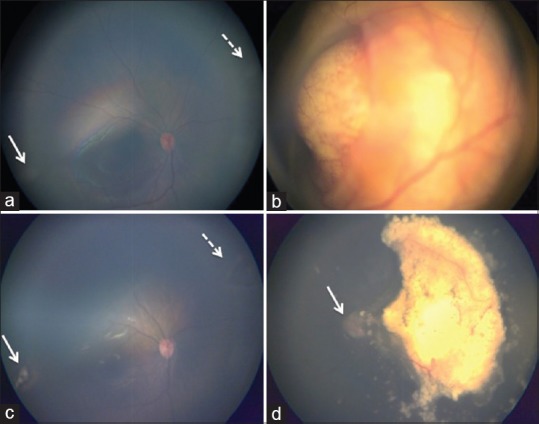
Figure 2.

Color fundus photograph of both eyes pre-treatment and post-treatment. Pre- treatment a and b, (a) RB Group A - Shows multiple small retinal lesions in the inferotemporal (white arrow) and superonasal equator (white dotted arrow). Optic disc and fovea is within normal limits. (b) RB Group D- Large solitary whitish retinal mass overhanging the optic disc located at the macula. There is total retinal detachment with diffuse subretinal seeds. Post-treatment c and d, (c) shows complete regression of inferotemporal (white arrow) and superonasal (white dotted arrow) tumors with focal laser therapy. (d) following 2 sessions of selective ophthalmic artery intra-arterial chemotherapy, tumor was completely regressed (Type-1). Subretinal fluid has completely resolved and the optic disc is visualized. (white arrow)
