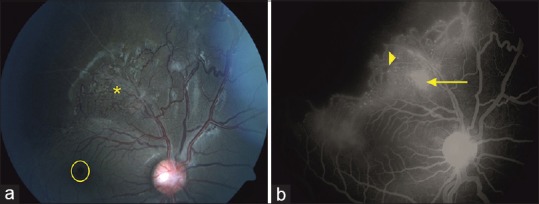
Figure 2.

Fundus picture at presentation showing gliosis over the disc, macular pseudohole (yellow circle) with superotemporal neovascularization (asterisk) and avascular retina (a). FFA at presentation shows the small FAZ, pin point leaks at the site of aneurysms (arrowhead), and leakage at the site of neovascularization with extensive CNP superotemporally (b)
