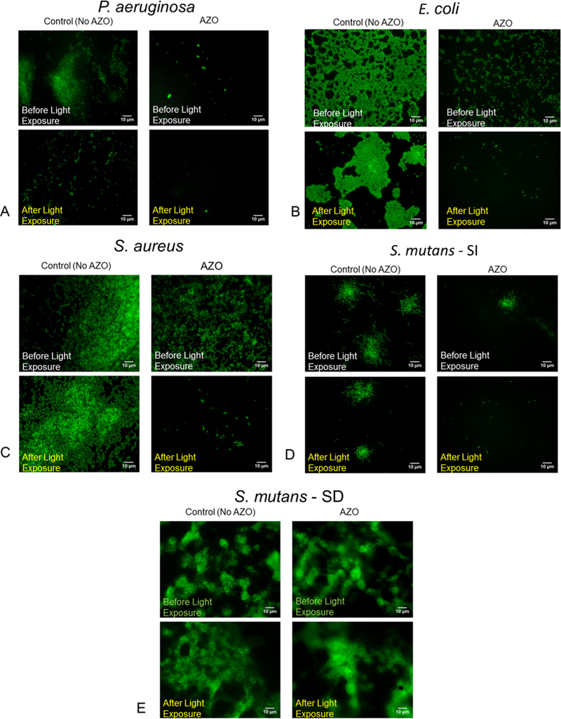
Figure 3.

Biofilms on the surface of the AZO substrates and the control substrates (No AZO) were exposed to light from a dental lamp (3 M Elipar DeepCure-S LED Curing Light) to initiate the fluidization effect and subsequently gently washed in PBS to remove the detached bacteria from the biofilm. This process was repeated 3 times for each sample. A live–dead stain before and after the light exposures and subsequent washes captures the ability of the photofluidization effect to remove bacterial biofilms from 4 different biofilms (3A–D). Our approach was not successful in removing biofilms formed via Streptococcus mutans in the presence of sucrose (3E).
