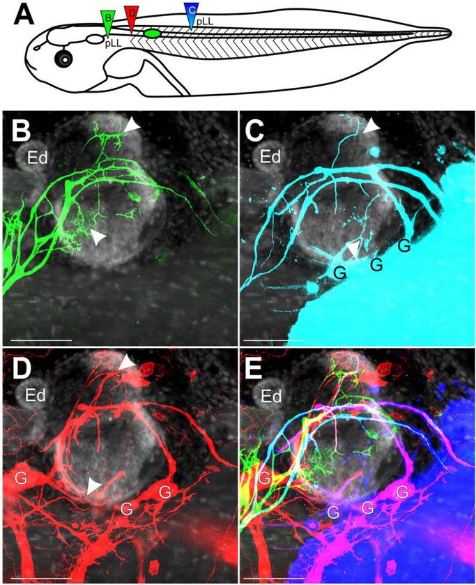
Figure 6.
Inner Ear Afferent Fasciculation with Lateral Line. (A) Schematic of dye placement for the different transplants. (B) Lateral view of afferents of the pLL projecting over and into the ear (green, dye injection B in panel A). The ear was transplanted adjacent to the spinal cord at stage 32–36. No ganglia were labeled. (C) Lateral view of pLL and inner ear afferents from a caudal dye injection into caudal portion of the pLL nerve (cyan, dye injection C in panel A). G, ganglia. (D) Lateral view of inner ear afferents from a spinal cord injection rostral to the transplanted ear (red, dye injection D in panel A) G, ganglia. (E) Merge of B-D. Cyan of panel C has been replaced by blue. Panels B–E are counterstained for nuclei marker Hoechst (gray). Arrowheads indicate areas of innervation of the inner ear. Scale bars are 100 μm. Endolymphatic duct, Ed. [Colour figure can be viewed at wileyonlinelibrary.com]

