
Fig. 5 |. Computational pathology tasks for machine learning applications.
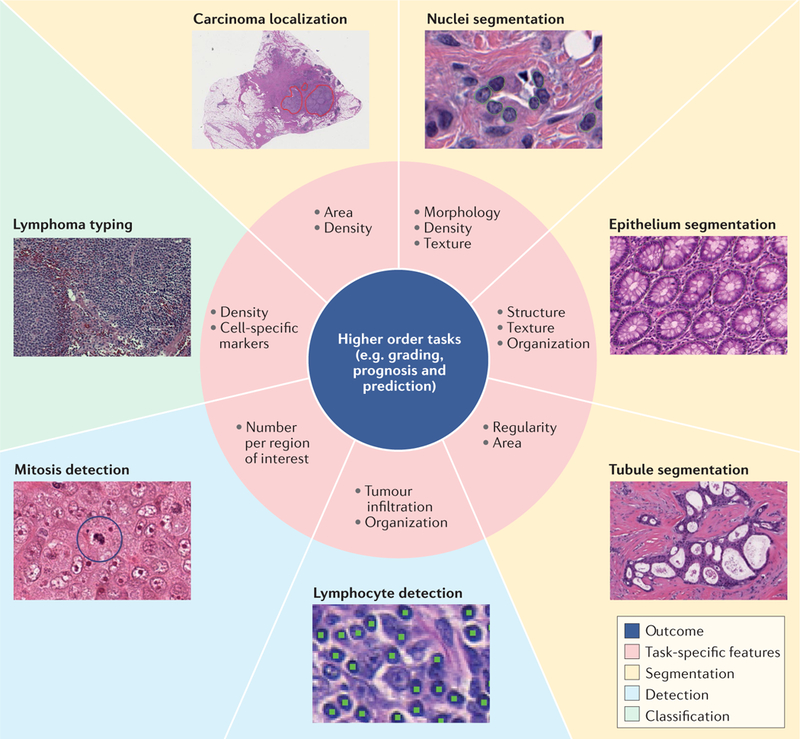
Deep learning frameworks can replace traditional handcrafted features in several basic pathology image-recognition tasks (such as segmentation of nuclei, epitheLia or tubules, lymphocyte detection, mitosis detection or classification of tumours) using image segmentation (yellow background), detection of specific features (blue background) or detection of a set of features used for classification (green background). Recognition is based on the task-specific features shown in the pink regions and can lead to more accurate prognosis or prediction of disease.