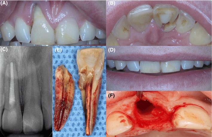
Figure 1.

Esthetic and radiographic overview of the failing right upper central incisor. A and B, clearly shows the hard and soft tissue deficiency and the fracture line, respectively. C, shows the radiologic assessment of failing tooth number 11 and fracture line. The patient's smile line was not visibly affected by the tissue deficiency. D, shows the patient's smile line. E, shows the extracted tooth, and F, shows both the hard and soft tissue deficiencies after tooth extraction
