
Figure 2.
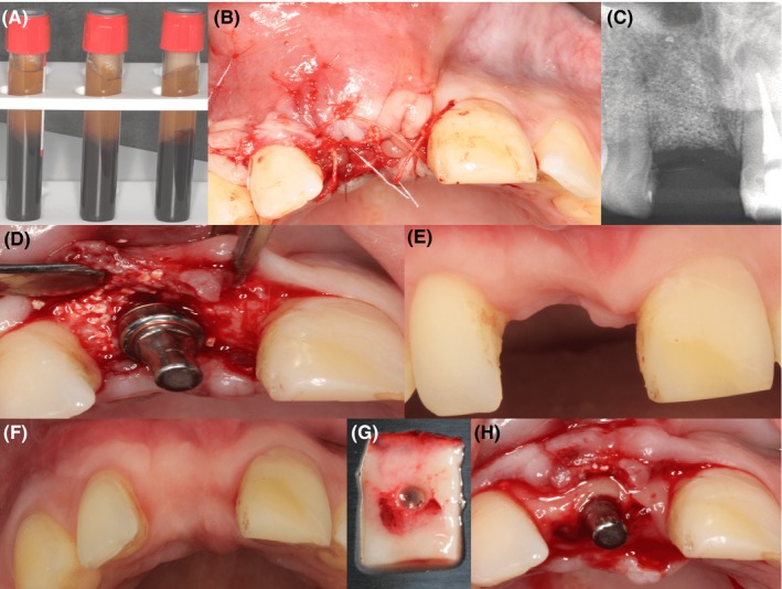
Preparation and the use of the PRF membrane. A, shows the PRF clot obtained after centrifugation. B, shows PRF membrane inserted into the cavity. C, shows the radiologic assessment of deficiency at 3 months after augmentation. D, shows implant placement with immediate temporary abutment. E and F, shows buccal deficiency after the buildup and occlusal appearance after buildup, respectively. G, shows the punched PRF membrane. H, shows PRF membrane placed in the buccal/palatal envelope