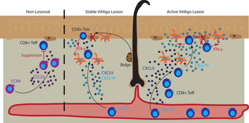
Figure 2. Chemokines promote T cell localization in the skin during vitiligo.

Left panel represents control of lesions by Tregs that naturally occupy the skin and suppress T effector cells. Tregs can be recruited via CCL22/CCR4 interactions (purple). Middle panel represents a stable vitiligo lesion in which resident memory T cells (Trm) are retained within the epidermis and maintain depigmentation. Melanocytes (brown) migrate out of the bulge of hair follicles and are recognized by Trm that secrete IFN-γ (red arrows), CXCL9 (dark blue), and CXCL10 (light blue) to recruit additional effector and central memory T cells to kill (large red X) new emigrants. Dermal antigen presenting cells (APCs) can respond to Trm IFN-γ and aid T cell recruitment through CXCL9 and CXCL10 release. Right panel represents an active vitiligo lesion in which high amounts of CXCL9, produced by dermal APCs, serve to recruit T cells to the skin, and CXCL10, produced by Langerhans cells (epidermal APCs) and keratinocytes (lighter brown cells in the epidermis), serves to facilitate localization up to the epidermis and destruction of melanocytes.
