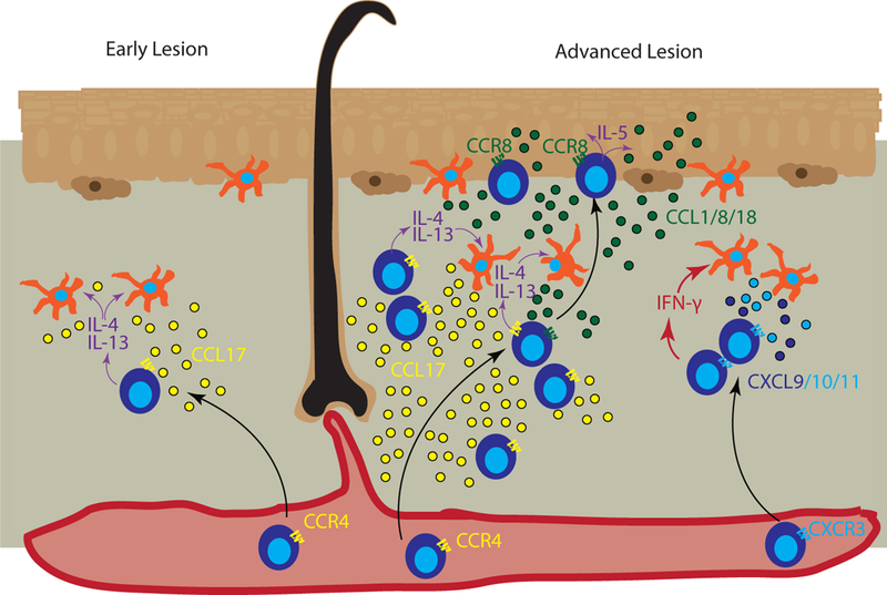
Figure 3. Chemokines promote T cell accumulation in atopic dermatitis.

Left panel represents early or developing atopic dermatitis (AD) lesions, in which CCR4+ T cells migrate into the skin and secrete type 2 cytokines IL-4/IL-13 (purple arrows), which act on skin antigen presenting cells (APCs) and other cells that produce CCL17 (yellow) to recruit additional CCR4+ T cells. Right panel represents advanced AD, in which IL-4-activated APCs secrete CCR4 ligands, predominantly CCL17, to recruit additional T cells. Keratinocytes in lesional skin secrete CCL1/CCL8/CCL18 (green dots) to recruit CCR8-expressing T cells into the epidermis. T cells in the epidermal compartment continue secreting IL-5 (purple arrows), which induces keratinocytes to secrete more CCL1/CCL8/CCL18, creating a positive feedback loop. In advanced lesions, additional inflammatory signals are increased including Type 1-skewed T cells that secrete IFN-γ (red arrows), which induces CXCL9/CXCL10 (dark and light blue dots) production by APCs to recruit additional CXCR3+ T cells, resulting in a mixed type 1/type 2 chemokine profile seen in chronic exacerbated AD.
